Recent development in Rapid Microbiology Methods
Posted: 29 September 2008 | Steve O’Hara, Director of Microbiology, Acolyte Biomedica Ltd | No comments yet
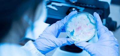
Microbiology is the scientific study of micro-organisms and includes many sub disciplines like bacteriology, virology, mycology, and parasitology: all characterized by the study of organisms too small to be seen by the naked eye. This defining aspect has determined the focus of microbial research over the last century: the need to selectively concentrate or amplify micro-organisms or their components to detect their presence. Earlier efforts into microbial detection involved microscopy, where light was transmitted through or reflected from the sample through lenses to magnify the view of the sample and determine the presence of micro-organisms. The limitations of light microscopy at the time prevented the study of living micro-organisms and it wasn’t until Pasteur (1822-1895) developed techniques to disprove the theory of spontaneous generation, and Robert Koch (1843-1910) developed his ‘postulates’ proving that diseases were caused by specific bacteria, was the era of microbial culture born.
Microbiology is the scientific study of micro-organisms and includes many sub disciplines like bacteriology, virology, mycology, and parasitology: all characterized by the study of organisms too small to be seen by the naked eye. This defining aspect has determined the focus of microbial research over the last century: the need to selectively concentrate or amplify micro-organisms or their components to detect their presence. Earlier efforts into microbial detection involved microscopy, where light was transmitted through or reflected from the sample through lenses to magnify the view of the sample and determine the presence of micro-organisms. The limitations of light microscopy at the time prevented the study of living micro-organisms and it wasn’t until Pasteur (1822-1895) developed techniques to disprove the theory of spontaneous generation, and Robert Koch (1843-1910) developed his ‘postulates’ proving that diseases were caused by specific bacteria, was the era of microbial culture born.
Microbiology is the scientific study of micro-organisms and includes many sub disciplines like bacteriology, virology, mycology, and parasitology: all characterized by the study of organisms too small to be seen by the naked eye. This defining aspect has determined the focus of microbial research over the last century: the need to selectively concentrate or amplify micro-organisms or their components to detect their presence. Earlier efforts into microbial detection involved microscopy, where light was transmitted through or reflected from the sample through lenses to magnify the view of the sample and determine the presence of micro-organisms. The limitations of light microscopy at the time prevented the study of living micro-organisms and it wasn’t until Pasteur (1822-1895) developed techniques to disprove the theory of spontaneous generation, and Robert Koch (1843-1910) developed his ‘postulates’ proving that diseases were caused by specific bacteria, was the era of microbial culture born.
The ease of use, low cost, and general applicability of culture means that despite scientific advances in the field of microbiology it remains the mainstay of microbial detection throughout the world today. Despite this dominance, with revenues from microbial culture roughly double the combined revenues of other techniques, recent industry reports suggest that microbiology culture is in the decline stage of its life cycle, whilst other techniques like immunoassay, and molecular techniques are in mature and growth phases respectively. This shift in microbiological methods has been driven by the introduction of enabling technologies which obviate, or at the very least mitigate, the need for biological amplification. These new approaches generally work by removing the need to detect the whole organism by amplifying cellular components, like nucleic acid, or using signal amplification to enhance detection capability.
The aim of this article is to review recent advances in microbiology and how these are impacting on the future microbiology landscape. As with Pasteur and Koch many technologies and advances mentioned in this article have been developed for clinical applications, or the need to detect and identify biothreat agents. However, these approaches have generic applicability across the science of microbiology and it is for each of us to consider the implications of these changes in our own discipline.
Developments in Rapid Microbiology Methods
There are many different ways to categorise the different types of technologies that are starting to shape the future microbiology landscape. One of the simplest and most fundamental approaches is to examine the methods used to concentrate and/or amplify micro-organisms and/or their components. These are presented in isolation but in practice are often used in combination to accentuate the affect and accelerate detection times.
Growth based amplification technologies
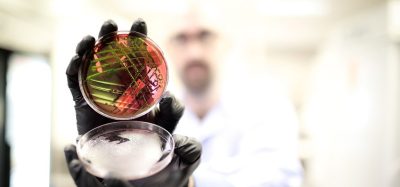
Substrate utilisation and biochemical tests are an established method for distinguishing bacterial species in pure cultures and have also been used in selective culture media, often coupled with pH indicators. The last 10 years has seen an expansion in the use of chromogenic substrates in growth media for the rapid detection of bacteria in food, clinical, and environmental samples. These media exploit chromogenic substrates that can be hydrolysed by specific bacterial species to yield coloured chromogens that remain restricted to bacterial colonies and do not defuse in agar. This allows isolation and detection of target pathogens within mixed cultures. Whilst chromogenic agars improve the ability to differentiate between bacterial types they are non selective and selective agents such as antibiotics often needed to be added to the media in samples from ‘dirty’ sites. The addition of selective agents often impacts on growth rates of target as well as non target bacteria leading to delays in the time to result. A more recent development which has greatly improved agar selectivity from ‘dirty’ samples has been the introduction of inhibigens. When added to a culture medium, these unique molecules provide highly specific selectivity as only organisms with the required uptake mechanism and specific enzyme to cleave the inhibitor-substrate complex are inhibited. This approach allows very specific inhibition of competing, non-target organisms with little or no impact on the target organism, even when cells are stressed. This means that recovery of specific organisms is improved in two ways: by reducing the growth of competing flora and by minimising exposure to potentially inhibitory components.
Enhancing the differential and selective properties of microbial culture media has improved culture media ease of use and had a small impact on time to result by reducing the number of ‘breakthrough colonies’ that have to be investigated further. In contrast the introduction of automated systems utilising optical, fluorescent or luminescence readers has helped shorten the time to detect growth changes when compared to visual or enzymic reactions. Examples include the Microscan (Dade Behring), Vitek (BioMerieux), Phoenix (Becton Dickinson) and BacLite System (3M Healthcare). These systems use sensitive detection technologies to replace the traditional visual end point read reducing operator subjectivity and time to result. They are often supplemented with advanced interpretative software to reduce background noise and smooth out erroneous data points, improving test accuracy and reproducibility.
Another approach used to reduce detection times in microbiology is to concentrate organisms from a large to small volume, effectively enhancing test sensitivity and potentially reducing the time to result. In addition to concentrating cells many of these approaches extract the organism from the specimen matrix which removes growth and assay inhibitors. Common examples of this type of approach include filtration and centrifugation. Lysis centrifugation is a technique used in clinical laboratories to concentrate small numbers of bacteria after lysis of blood components in blood samples. Buoyant density centrifugation has been used in the food industry to concentrate bacteria in food samples. Another separation and concentration technology which is widely used in many scientific disciplines and the workhorse of many high throughput automated systems such as the BEADS (Biodetection Enabling Analyte Delivery System) platform is immunomagnetic separation. In this approach the sample is incubated with paramagnetic beads tagged with antibodies to the target organism. The antibody binds to the target which is then is extracted from the sample by the introduction of a magnetic field which attracts the magnetic bead-antibody-antigen complex. This approach has been used to separate organisms from human fluids, food, and water, and is increasingly being used to automated nucleic acid extraction. A less known example of cell technologies based on cell concentration techniques include diolectrophoresis which uses the intrinsic dielectric properties of biological particles (e.g. mammalian cells, bacteria, viruses and other non-cellular biological agents) to separate cells in a non uniform electric field.
Immunological Testing
The introduction of monoclonal antibodies with high target specificity has led to a number of advances in rapid microbiology techniques. Agglutination tests employing monoclonal antibody bound latex beads have aided the rapid and specific identification of a number of organisms such as Staphylococcus aureus and Streptococcus pneumoniae from bacterial cultures. However, these tests tend to lack sensitivity which has limited their usefulness from analytical samples where microbial numbers may be low. Concentration techniques such as ultrasound can enhance test sensitivity up to 64 fold by increasing the contact between antibody coated microparticles suspended in the sound field enhancing the rate of antigen antibody interaction. Despite these sensitivity improvements agglutination tests are largely used for culture confirmation.
Immunoassays consisting of essentially antibody based capture; a method to separate bound and unbound antibody antigen complexes and a detection devise are common in clinical, pharmaceutical, and food microbiology. Immunoassay performance is dependent upon two major factors: the efficiency of the antibody antigen formation and the sensitivity of the detection technology. Historically, immunoassays have lacked sensitivity, with detection limits in the region of 105 cfu/ml, and been used to detect a single analyte. However, advances in assay design, matrix format, and detection methodologies (e.g. fluorescence, chemiluminescence), have resulted in the development of commercial multiplex immunoassays with good performance characteristics. Whilst many formats exist most are variations of solid support platforms or lateral flow devices. The most commonly used solid support platform is the Enzyme Linked Imunosorbent Assay (ELISA) in which one component of the assay, either the antigen or the antibody is immobilised on a solid support. Recent developments of solid phase platforms include coating antibodies targeted at different analytes onto different coloured polystyrene beads allowing detection of multiple analyse in a single multiplex assay (e.g Luminex xMAP). As each bead is spectrally unique laser excitation leads to signal generation proportional to the target concentration in the samples. Using this approach it is theoretically possible to detect multiple analytes (100) in samples.
Lateral flow tests are used widely across microbiology and work by binding antibodies or some other capture agent, such as microphages, to a detection marker. These detection markers are usually very small pieces of gold, called colloidal gold, or small coloured plastic beads. When a liquid sample wicks across the paper strip or membrane it encounters the antibody/marker combination. If the antigen of concern is present, antibody–antigen complexes form which continue to flow across the membrane until the complex is captured by a second immobilized antispecies antibody. If enough of the markers are present in the test region, a visible line will appear. These tests are best characterised by home use pregnancy tests and their speed and ease of use have facilitated widespread adoption. Lateral flow tests have received increased interest with the bioterrorism threat with tests being developed for B. anthracis, Y.pestis, C.botulinum, and other potential threat agents.
Biosensors
Over the past two decades, enormous activity has taken place in the field of sensor technology which couple signal amplification technologies with a sensors response to the presence of microbes or their products. Biosensors, in particular, have attracted considerable attention because of their potential for the detection of multiple analytes, miniaturisation and point of care use. As there name suggests Biosensors utilise biological sensing elements in proximity to the signal transducer with an array of signal collecting and pattern recognition software devices. Biological sensing generally use some form of immunological or probes based target recognition to detect microbes (direct sensing) or their products (indirect sensing). Increasingly other ligands, such as aptamers, which exploit shape based, rather than sequence based recognition, are being used. Aptamers are small (i.e., 40 to 100 bases), synthetic oligonucleotides that can specifically recognize and bind to virtually any kind of target including whole cells and toxins with a higher degree of specificity than antibody preparations and have been used to detect a range of bacteria and their toxins.
Probably the best known example of an indirect biosensor is the electronic nose which detects the volatile organics compounds and gases produced by growing bacteria. The technique has been used for the detection of bacterial vaginosis and the presence of bacteria in urine with limited success. Biochemical biosensors like the electronic nose are indirect sensors in that they detect biological products of the target. As these products vary depending on nutritional state, environmental conditions and the co-presence of other organisms these sensors often require complex pattern recognition software and tend to lack specificity.
Despite good analytical performance, low-cost instrumentation, minimal sample preparation, small sample size, and fast response times the potential of biosensors remains unfulfilled in microbiology. However, the success of biosensors in glucose monitoring, an application which makes up 90% of this field, suggest they hold promise for the future. One of the principal challenges remaining in this field is that many biosensor devices lack storage and operational stability because they are based on a fragile biological recognition element: an enzyme or antibody. An emerging technology called molecular imprinting, however, could provide an alternative. This technique leads to highly stable synthetic polymers that possess selective molecular recognition properties because of recognition sites within the polymer matrix that are complementary to the analyte in the shape and positioning of functional groups. Some of these polymers have high selectivity’s and affinity constants, comparable with naturally occurring recognition systems such as monoclonal antibodies or receptors, which make them especially suitable as constituents in analytical sensors.
Molecular methods
In the last decade we have witnessed techniques for amplifying and detecting nucleic acid move from research into main stream microbiology. Generally, molecular microbiology methods can be broken down into hybridisation and amplification approaches.
Hybridisation techniques are considered the origin of molecular microbiological methods and use nucleic acid probes with genetic sequences complementary to the target organism and radioactive, fluorescent, or enzymic labels as target signals. Hybridisation methods usually require large amounts of starting target nucleic acid which means that these methods often lack sensitivity and tend to be used to confirm amplification products (e.g. from PCR) or for the identification of culture isolates. Fluorescence in situ hybridization (FISH) is good example of a hybridisation technique in which fluorescently labelled nucleic acid or peptide nucleic acid (PNA) probes are hybridized to complementary targets located with cells and viewed using fluorescent microscopy. Earlier incarnations of this technique often suffered from limited sensitivity due to low target copy number or target inaccessibility but recent developments using rRNA targets and multiply labelled polynucleotide probes (poly FSH) have helped increased signal intensity and rekindled interest in this technique.
In analytical settings most samples for analysis rarely contain sufficient nucleic acid to allow direct detection by hybridisation methods and target nucleic acid therefore has to be increased by Nucleic Acid Amplification Techniques (NAAT). Amplifying a target sequence can be achieved by a host of nucleic acid amplification technologies including the Polymerase Chain Reaction (PCR), Nucleic Acid Sequence Based Amplification (NASBA), and Transcription mediated amplification (TMA) to name just a few. Alternatively, a probe that has been recognised and annealed to a target sequence can be amplified as in the Ligase Chain Reaction (LCR). The main advantage of nucleic acid amplification methods is rapid amplification of a specific target sequence allowing analytical sensitivity to be increased in a short time period, typically a couple of hours. Multiple targets can often be amplified simultaneously (e.g. multiplex PCR), to look for multiple organisms in a single sample or consecutively (e.g. nested PCR), to further enhance test performance. In early years the promise of molecular amplification tests remained largely unfulfilled due to complexities of sample preparation and the techniques used to detect amplified products. The introduction of Real time PCR, in which the nucleic acid is quantified after each round of amplification by the use of intercalating dyes or reporter molecules which signal when hybridized with a complementary DNA, reduced the need for complex and time consuming analysis of amplified products and removed a major barrier to uptake in analytical settings. In many areas of microbiology nucleic acid based tests gradually are replacing or complementing culture-based, biochemical, and immunological assays. Recent improvements in detection technologies have paved the way for the development of multiparameter assays while the introduction of closed-tube real-time polymerase chain reaction systems has resulted in the development of rapid microbial methods with a reduced contamination risk.
Despite the undoubted promise of molecular methods a number of challenges remain. These include the potential for contamination and false positive results, the high technical skill requirement required in some test protocols, lack of standardization or validation of many assays, the complexity of interpreting results, and the relatively high cost of instrumentation and reagents. Whilst these challenges have limited broad based acceptance of molecular methods across all areas of microbiology there is an increasing realisation that molecular tests will have a growing role to play in the future.
Conclusions
This brief overview of rapid microbiological methods in microbiology attempts to discuss recent advances in microbiology. Rather than simply list a plethora of new technologies it has attempted to identify common development themes. Most of the development approaches discussed are based around removing the need to detect the whole organism by concentrating or amplifying cellular components, like nucleic acid, or using signal amplification to enhance detection capability. Many of these technologies have been around for a number of years but have gained a resurgence of interest due to technological changes often unrelated to the fundamental science. For example, PCR was discovered by Kary Mullis in 1983 but it wasn’t until the complexities of sample preparation and techniques to detect amplified products were developed did the technique start to fulfil some of its promise. Herein lies the crux of rapid microbiology testing. There are many technologies which have the potential to accelerate testing times and change the face of microbiology. However, predicting the future on the basis of technological and scientific merit alone is fraught with danger. To gain widespread acceptance these technologies have to be configured to meet broader user needs such as cost and convenience. In today’s world its not just about scientific merit but user and application fit with different users having different needs depending on their microbiology discipline and situation. In truth there is unlikely to be an ideal technology for all situations.