

Biomarker breakthrough could improve Parkinson’s treatment
Posted: 16 August 2016 | | No comments yet
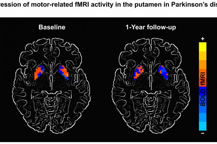
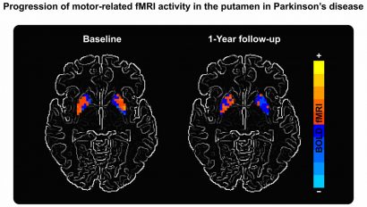
Scientists have found that using magnetic resonance imaging reveals areas where Parkinson’s disease causes progressive decline in brain activity, a biomarker discovery which will help to evaluate new experimental treatments to slow or stop the disease’s progression…
Scientists have found that using magnetic resonance imaging reveals areas where Parkinson’s disease causes progressive decline in brain activity, a biomarker discovery which will help to evaluate new experimental treatments to slow or stop the disease’s progression.
While current treatments focus on controlling symptoms, biomarker provides a quantifiable way to measure how medications address not just symptoms, but the neurological changes behind them.
Previous studies have used imaging techniques that require the injection of a drug that crosses the blood-brain barrier but this is the first “non-invasive treatment and it’s much less expensive”, according to David Vaillancourt, PhD Department of Applied Physiology and Kinesiology, University of Florida.
Magnetic resonance imaging was used to evaluate five areas of the brain, key to movement and balance. A year after the baseline study, the 46 Parkinson’s patients in the study showed declining function in two areas: the primary motor cortex and putamen.
Parkinson’s-related disorders evaluated in the study also showed declines with the 13 subjects with multiple system atrophy had reduced activity in three of the five areas, while the 19 with progressive supranuclear palsy showed declines in all five areas. The brain activity of the 34 healthy control subjects did not change.
“For decades, the field has been searching for an effective biomarker for Parkinson’s disease” as they “can be used to monitor the progression”, said Debra Babcock, MD PhD, NIH’s National Institute of Neurological Disorders and Stroke.
An earlier study documented Parkinson’s development via MRI, showing an increase in unconstrained fluid in an area of the brain called the substania nigra. An NIH-funded study beginning in November will use both biomarkers to test if a drug approved for symptom relief can slow or stop the progressive degeneration.
