


Baldur Sveinbjørnsson, Chief Scientific Officer of Lytix Biopharma, discusses how the clinical insights from the Phase II NeoLIPA trial of neoadjuvant intratumoral therapy could inform the future of neoadjuvant trial design.
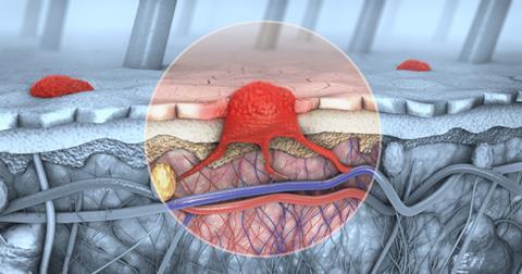
For decades, intratumoural (IT) oncology was primarily regarded as a local intervention, used to debulk accessible lesions, manage cutaneous recurrences or provide palliative control of symptomatic disease. Today, accumulating clinical and translational data indicate that the true value of IT approaches lies in their capacity to generate systemic, tumour‑specific immune responses that extend far beyond the injected lesion, with direct implications for neoadjuvant trial design and combination immunotherapy strategies.
In melanoma, this shift is already visible in routine practice. Neoadjuvant (pre-surgical) immunotherapy is rapidly emerging as best practice in clinical stage III disease, with immune checkpoint inhibitors delivering high pathological response rates and improved recurrence‑free survival.1-3 Early trials with intratumoural agents, including oncolytic viruses and oncolytic peptides, suggest that directly treating primary and in‑transit lesions can further amplify these neoadjuvant benefits by broadening T‑cell clonality and deepening local microenvironment remodelling at the site of disease.4-6
Within this evolving landscape, the neoadjuvant setting is becoming a biologically powerful and operationally manageable context in which to maximise the potential of intratumoural therapy. Treating an intact primary tumour before resection allows intratumoural agents to remodel the local microenvironment and prime systemic immunity, with a primary objective of lowering post‑operative recurrence risk.
Why the intact melanoma tumour matters in the neoadjuvant setting
In patients with resectable stage III melanoma and in‑transit disease, intervening prior to lymph node dissection or wide excision confers distinct biological advantages that are attenuated or lost once the primary and regional disease are removed. These advantages underpin the concept of the primary tumour as a patient‑specific in vivo vaccine site.
- · Comprehensive antigen reservoir: The primary tumour contains the full spectrum of clonal and sub‑clonal mutations unique to each patient, creating a concentrated in situ source of neoantigens for immune priming. Local cytolysis in this setting releases a broad repertoire of patient‑specific antigens, enabling multiclonal T‑cell activation that can, in principle, target both the injected lesion and occult micrometastatic disease.
- · Intact lymphatic architecture: Effective immune priming depends on a functional lymphatic system. In the neoadjuvant window, regional lymphatics and draining lymph nodes remain largely undisturbed by surgery or radiation, enabling antigen‑presenting cells to traffic efficiently, present neoantigens and activate T cells in a physiologically intact environment.
- · Less exhausted immunity: Earlier‑stage patients generally have more preserved immune function and lower levels of T‑cell exhaustion than heavily treated, late‑stage populations, providing a more responsive substrate for immunogenic intratumoural interventions.
When a cytolytic intratumoural agent is administered into this environment, the objective is not simply local tumour destruction but the initiation of a coordinated immunogenic cascade. Rapid tumour-cell death releases neoantigens alongside damage‑associated molecular patterns (DAMPs) and other danger signals, driving recruitment and maturation of dendritic cells, trafficking to lymph nodes and priming of effector and memory T‑cell populations. In principle, these T cells can recognise both the injected lesion and undetected microscopic metastases, transforming the primary tumour into an in vivo (in situ?) vaccine site for multiclonal systemic antitumour immunity.7,8
Tumour heterogeneity and immunosuppressive microenvironments
Tumour heterogeneity remains a central limitation for single‑target systemic therapies and single‑pathway inhibitors, which often fail to control diverse sub‑clonal populations within the same lesion. Intratumoural therapies with an immunogenic, cytolytic mode of action may partially circumvent this barrier by exposing the immune system to a wide spectrum of tumour antigens simultaneously at the site of primary disease, promoting multiclonal T‑cell responses.
Clinical evaluations in late‑stage settings have shown that local intratumoural treatment can expand many distinct T‑cell clones, accompanied by observable immune effects in distant, non‑injected lesions (abscopal effects).9,10 While these observations require confirmation across tumour types and larger controlled datasets, they support the concept that multiclonal activation may be particularly relevant for immune‑excluded or immune suppressive tumours, where checkpoint inhibition alone is insufficient.
Ruxotemitide and overcoming checkpoint resistance
Neoadjuvant intratumoural approaches are already being explored in melanoma. Preclinical and early clinical work with oncolytic peptides such as ruxotemitide (LTX‑315) further support the concept that local cytolysis can convert poorly immunogenic tumours into ‘immunoactive’ lesions characterised by dense CD8+ T‑cell infiltration, potentially overcoming resistance in patients who have failed multiple lines of systemic checkpoint inhibitors.11,12
Systemic immune checkpoint inhibitors (ICIs) have transformed outcomes in several malignancies, but their activity depends on a pre‑existing or inducible antitumour immune response. In desert‑like or highly excluded phenotypes, releasing inhibitory signals via ICIs is insufficient if T cells cannot traffic into or persist within the tumour microenvironment. Intratumoural therapy can serve as a mechanistically complementary approach: by inducing local tumour lysis and inflammation, enhancing antigen release and modifying stromal and vascular features, it can create a more permissive microenvironment in which systemic immunotherapies and rational combinations are more likely to succeed, including in ICI‑refractory melanoma.13
Shifting objectives and pathological response
Volume reduction and improved resectability are practical benefits of neoadjuvant intratumoural therapy, but they should not be considered the primary endpoint for this modality. For neoadjuvant intratumoural immunotherapy to be meaningfully differentiated, the central goal must be long‑term disease control, reflected in pathological response rates and event‑free survival.
A clinically relevant hierarchy of objectives in this setting includes:
- · Induction of systemic immunity: Demonstrating that a localised intratumoural intervention can reliably trigger durable, systemic tumour-specific responses, as reflected by changes in peripheral T‑cell repertoires and functional assays.
- · Enhancement of pCR and MPR rates. In melanoma, neoadjuvant checkpoint inhibitor studies have shown that major pathological responses correlate strongly with recurrence‑free survival; intratumoural strategies should be designed to at least match, and ideally exceed, these benchmarks, positioning high pathological response rates as a key readout for future neoadjuvant trial design.2,3
- · Improvement in event‑free survival (EFS). For high‑risk stage III melanoma, linking intratumoural‑driven pathological responses to durable control of nodal and distant relapse will be a critical measure of clinical value and will inform how intratumoural agents are integrated into standard neoadjuvant pathways.
- · Informing adjuvant strategy. Using the neoadjuvant window to collect high‑quality translational data – spanning tissue, blood and digital pathology – can guide adaptive adjuvant decision making based on real‑time microenvironmental remodelling, an approach that aligns closely with the priorities of pharmaceutical development teams.
Anchoring trial design in these endpoints positions intratumoural therapy as a data‑driven, immunologically purposeful component of care”
Anchoring trial design in these endpoints positions intratumoural therapy as a data‑driven, immunologically purposeful component of care.
Timing and coordination in neoadjuvant protocols
Operationalising neoadjuvant intratumoural therapy requires close collaboration among medical oncologists, surgeons, radiation oncologists and translational research teams. The key challenge is temporal: allowing sufficient time for antigen release, dendritic cell trafficking, T‑cell priming and peripheral infiltration, while preserving an appropriate surgical window and minimising risk.
Different intratumoural modalities operate on distinct kinetic timelines
Oncolytic molecules
Directly cytolytic agents often act rapidly, causing membrane disruption and antigen release over hours to days.
Oncolytic viruses
Viral platforms depend on uptake, intracellular replication and subsequent lysis, producing a more extended and potentially cyclical pattern of antigen exposure.10
Dose, schedule and sequencing with surgery and systemic therapies must be intentionally co‑designed around these biological kinetics. Protocols should integrate predefined imaging and biospecimen time points to capture the dynamics of immune activation without compromising surgical standards or patient safety.
Building a data‑rich pathway to mainstream adoption
To bring intratumoural therapy into the mainstream neoadjuvant paradigm over the next several years, trial architecture and translational infrastructure will need to evolve beyond the ‘single‑procedure’ mindset. Instead, IT interventions should be embedded within comprehensive studies that couple optimised injection techniques with robust biomarker strategies.
NeoLIPA and future trial design
Melanoma is uniquely well suited to data‑rich neoadjuvant experimentation, given the accessibility of lesions for repeated biopsy, the existing evidence base correlating pathological response with long‑term outcomes and the availability of multiple active systemic immunotherapies.
Investigator‑initiated trials such as NeoLIPA (NCT06651151), an open‑label Phase II study in resectable stage III–IV melanoma evaluating intratumoural ruxotemitide in combination with pembrolizumab in the neoadjuvant setting, exemplify how intratumoural agents can be integrated into modern melanoma care while generating granular translational data. NeoLIPA also illustrates how high pathological response rates, if confirmed, could become pivotal endpoints for future neoadjuvant intratumoural trials.6
By systematically characterising how local intratumoural treatment reshapes both the tumour microenvironment and systemic immunity, it should be possible to refine patient selection, identify rational combinations with ICIs and other agents and extend this strategy beyond melanoma into additional solid tumours where neoadjuvant standards are still evolving – a prospect of clear interest to pharmaceutical developers.
About the author
Baldur Sveinbjørnsson is Chief Scientific Officer of Lytix Biopharma and has been involved in the company’s research activities since its inception. He received his PhD from the Faculty of Medicine at the University of Tromsø in 1998, where he studied mechanisms and mediators of immunomodulation in experimental tumours, and now focuses on developing oncolytic molecules for intratumoural administration, with particular interest in neoadjuvant strategies that link tumour destruction, antigen release and translational biomarker frameworks.
References
1. Awada G, Cascone T, van der Heijden MS,et al. Nature Cancer 2025, 6 (6), 967–987. DOI: 10.1038/s43018-025-00990-7.
2. Menzies AM, Amaria RN, Rozeman EA, et al. Nature Medicine 2021, 27 (2), 301–309. DOI: 10.1038/s41591-020-01188-3.
3. Rawson RV, Adhikari C, Bierman C, et al. Annals of Oncology 2021, 32 (6), 766–777. DOI: 10.1016/j.annonc.2021.03.006.
4. Blank CU, Lucas MW, Scolyer RA, et al. New England Journal of Medicine 2024, 391 (18), 1696–1708. DOI: doi:10.1056/NEJMoa2402604.
5. Ferrari M, Facchini BA, Ascierto PA, Sparano F. Expert Review of Anticancer Therapy 2025, 25 (4), 383–392. DOI: 10.1080/14737140.2025.2474182.
6. Oslo University Hospital. NeoLIPA: Neoadjuvant LTX-315 in combination with pembrolizumab in resectable stage III/IV melanoma.
7. Eike L, Yang N, Rekdal Ø, Sveinbjørnsson B. Oncotarget 2015, 6 (33), 34910–34923.
8. Li X-Q, Yamazaki T, He T, et al. Frontiers in Immunology 2024, Volume 15 - 2024. DOI: 10.3389/fimmu.2024.1332922.
9. Spicer J, Marabelle A, Baurain J-F, et al. Clinical Cancer Research 2021, 27 (10), 2755–2763. DOI: 10.1158/1078-0432.Ccr-20-3435.
10. Kaufman HL, Amatruda T, Reid T, et al. J Immunother Cancer 2016, 4, 12. DOI: 10.1186/s40425-016-0116-2.
11. Camilio KA, Wang MY, Nestvold J, et al. Cancer Research 2017, 77 (13_Supplement), 11–11. DOI: 10.1158/1538-7445.Am2017-11.
12. Yamazaki T, Pitt JM, Vétizou M, et al. Cell Death & Differentiation 2016, 23 (6), 1004–1015. DOI: 10.1038/cdd.2016.35.
13. Marabelle A, Andtbacka R, Harrington K, et al. Ann Oncol 2018, 29 (11), 2163–2174. DOI: 10.1093/annonc/mdy423.





No comments yet