Infrared thermography can detect pre-visual bacterial growth, shows study
Posted: 19 August 2021 | Hannah Balfour (European Pharmaceutical Review) | No comments yet
A new paper shows thermal imaging (infrared thermography) can detect E. coli and S. aureus bacteria after just six hours of incubation, long before it is visible to the human eye.
New research suggests laboratories and manufacturing facilities could implement infrared thermography, also known as thermal imaging, to detect metabolically active bacteria more rapidly – before they even become visible to the human eye.
Current methods for bacteria detection do not give results in real time, instead it may take many hours, or days, for samples to be analysed using the various bacterial culturing, molecular- and biochemical-based detection methods available. This has led to the development of rapid microbiological methods (RMMs).
Thermal imaging – which uses an infrared camera to detect heat – is one of these RMMs; however, its use in bacterial detection is in its infancy. In previous experiments thermal imaging has been able to detect Escherichia coli in liquid broth at levels as low as 120 colony forming units (CFU) per ml; Staphylococcus aureus, Vibrio chloerae, Vibrio mimicus, Proteus mirabilis and Pseudomonas aeruginosa in liquid broth; and E. coli on agar plates after only 11 hours of growth.
In a new paper published in The Journal of Applied Microbiology, researchers investigated infrared thermography as a potential non-invasive method of detecting bacterial growth. They used E. coli and Staphylococcus aureus on solid growth media in their experiments: streaking one half of the surface of an agar plate with bacteria from a single colony using an inoculating loop and the other half with a sterile loop to give to act as an in-built plate control. Other plates were streaked with sterile loops only to give an environmental control.
The plates were then incubated for six hours at 37℃ before being individually removed and thermal videos captured, then replaced in the incubator and imaged at 24 hours. Some of the plates were irradiated with ultraviolet (UV) light at 312nm for a minute before returning to the incubator for an hour. Additionally, 10μl of 0.1 mmol/L of 2,4-dinitrophenol (DNP) was added to eight locations on the streaked section of the plate and the plates incubated at 37℃ for a further 18 hours before the thermal video recording was repeated.
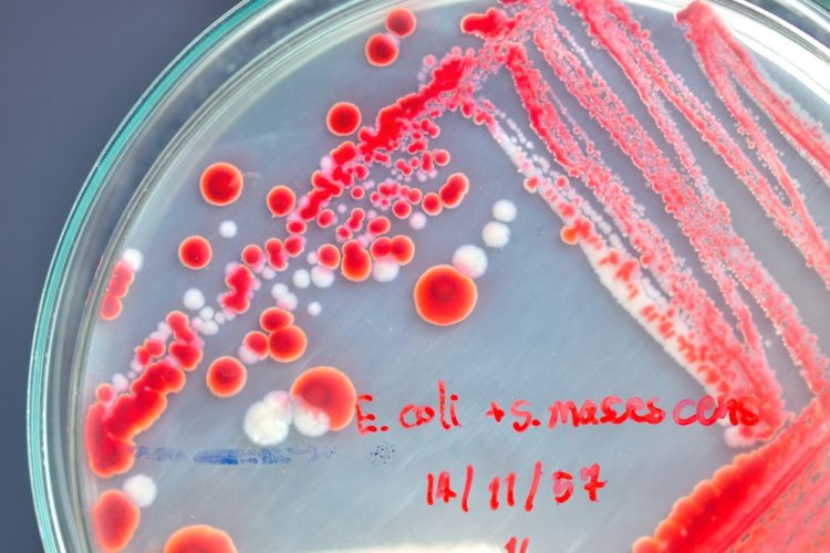
![Representative visual and thermal images of agar plates streaked with Escherichia coli and Staphylococcus aureus at 6 and 24 h of growth. E. coli and S. aureus were streaked onto the right-hand side of the agar plate and incubated at 37℃ for 24 h with visual (a) and thermal images (b) taken at 6 and 24 h. The dashed line indicates the midline of the plate dividing the plate into the areas with and without the bacterial growth. Thermal images were taken within 30 s of the plates leaving the incubator (n = 12) [Credit: Hunt B. et al, 2021].](https://www.europeanpharmaceuticalreview.com/wp-content/uploads/Infrared-thermography-375x192.jpg)
Representative visual and thermal images of agar plates. E. coli and S. aureus were streaked onto the right-hand side of the agar plate and incubated at 37℃ for 24 hours, with visual (a) and thermal images (b) taken at six and 24 hours. The dashed line indicates the midline of the plate dividing the plate into the areas with and without the bacterial growth. Thermal images were taken within 30 seconds of the plates leaving the incubator [Credit: Hunt B, et al, 2021].
The results show that infrared thermography can detect E. coli and S. aureus on solid growth media plates at six hours, before they are visually observable or detectable using standard visible spectrum photography. Moreover, it was demonstrated to be detecting viable, metabolically active bacteria, since a heat decrease was observed after treatment with UV light (to destroy the bacteria) and a more than four-fold heat increase was reported after incubation with DNP (which uncouples the electron transport chain, increasing mitochondrial activity, causing cells to produce more heat).
According to the researchers from Sheffield Hallam University, UK, thermal imaging can be practically applied in a laboratory, clinical or industrial setting since it is statistically robust, can be undertaken in situ and does not require specialised temperature control facilities.
The scientists did state that the temperature of a sample is likely to be influenced by the surrounding environment and dependent on the heat retention ability of the surface on which the bacterial cells are bound. Additionally, they cautioned that it may not be applicable in every instance since not all contaminating bacteria are metabolically active.
The researchers concluded that infrared thermography “may become an important methodology for the timely and straightforward detection of early-stage bacterial growth”.
Related topics
Analytical techniques, Environmental Monitoring, Microbial Detection, Microbiology, Rapid Microbiological Methods (RMMs), Technology