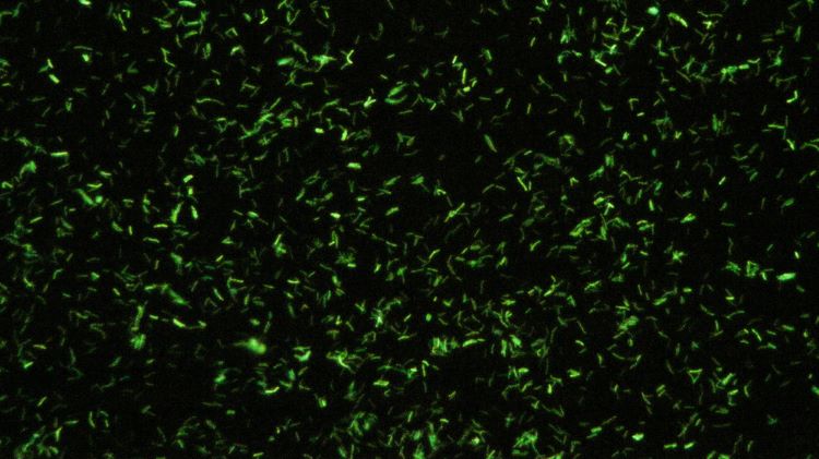
Innovative method could address a major limitation in fluorescence microscopy
Posted: 20 November 2024 | Catherine Eckford (European Pharmaceutical Review) | No comments yet
With fluorescence imaging an essential tool in noninvasive biomedical research, the microscopy advancement holds opportunity for progress in optical imaging, according to the paper.


Researchers in Israel have developed a way to obtain high-resolution imagery using megapixel fluorescence microscopy without needing specialist equipment.
The technique enables clear images of dense and challenging targets to be captured. It can correct computational scattering, which is one of the main limitations in fluorescence imaging, according to the paper that discusses the study findings.
The new method is compatible with standard microscopy setups and when used with established matrix-based techniques, it has widespread applicability, Weinberg et al. stated.
The framework is based on construction of a virtual reflection matrix via “a small number of measurements”. Specifically, “by treating each camera pixel as a random variable and calculating the covariance matrix [this allows] the applications of different scattering compensation techniques developed for reflection matrix analysis”.
Reconstruction of the megapixel-scale image was demonstrated with less than 150 widefield fluorescence-microscope frames, without any spatial light modulators or computationally intensive processing, Weinberg et al. explained.
Reconstruction of the megapixel-scale image was demonstrated with less than 150 widefield fluorescence-microscope frames, without any spatial light modulators or computationally intensive processing”
Overall, the method “is capable of considerably superior imaging in the case of nonsparse objects, while also requiring a substantially lower number of illuminations.”
Weinberg et al. elucidated in their paper that frames captured via their technique are “identical to those obtained in dynamic speckle illumination microscopy”. This is because like “in a conventional reflection matrix, the virtual fluorescence reflection matrix diagonal provides a “confocal” image”.
However, the researchers noted that “similar to conventional confocal microscopy, it is insufficient for imaging deep in scattering media”.
Developing the novel fluorescence microscopy imaging method
Weinberg et al. anticipated that future research “may use random illumination microscopy (RIM) variance matching to further improve the lateral resolution of the final images”.
This fluorescence microscopy research was supported by funding from the European Research Council and published in Science Advances.
Related topics
Data Analysis, Imaging, Microscopy, Research & Development (R&D), Technology


