New app replaces ultrasound to measure heart health
Posted: 6 September 2017 | Dr Zara Kassam (European Pharmaceutical Review) | No comments yet
Engineers have demonstrated that the camera on your smartphone can noninvasively provide detailed information about heart health…
Engineers have demonstrated that a smartphone camera app can noninvasively provide detailed information about heart health.
What used to require a 45-minute scan from an ultrasound machine can now be accomplished by simply holding your phone up to your neck for a minute or two.
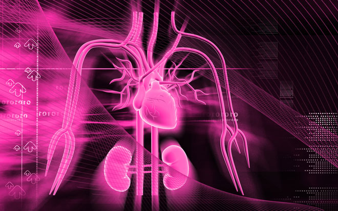
The team developed a technique that can infer the left ventricular ejection fraction (LVEF) of the heart by measuring the amount that the carotid artery displaces the skin of the neck as blood pumps through it.
LVEF is a key measure of heart health, one upon which physicians base diagnostic and therapeutic decisions.
“In a surprisingly short period of time, we were able to move from invention to the collection of validating clinical data,” says Dr Mory Gharib from Caltech University.
To test the app, clinical trials were conducted with 72 volunteers between the ages of 20 and 92 at an outpatient magnetic resonance imaging (MRI) facility. MRI is the gold standard in measuring LVEF but is seldom used clinically due to its high cost and limited availability.
LVEF is most commonly measured using an ultrasound machine during a procedure known as echocardiography. Echocardiography, however, requires a trained technician, an expensive ultrasound machine, and up to 45 minutes of a patient’s time.
To measure LVEF using the technique developed at Caltech, doctors simply held iPhones against the volunteers’ necks for one to two minutes. Afterwards, the volunteers immediately received an MRI examination, and data from both tests were compared. The measurements made by smartphone had a margin of error of ± 19.1 percent compared with those done in an MRI. By way of comparison, the margin of error for echocardiography is around ± 20.0 percent.
“What is exciting about this study is that it shows our technique is as accurate as echocardiography at estimating LVEF when both are compared to the gold standard of cardiac MRI. This has the potential to revolutionise how doctors and patients can screen for and monitor heart disease both in the U.S. and the developing world,” said Dr Gharib.
The app works because the walls of arteries are almost completely elastic–they expand and contract with each beat of the heart. That expanding and contracting can be measured and described as a waveform that encodes information about the heart. For the study, the team used an iPhone 5, but any smartphone with a camera will work.
Dr Gharib and his team are exploring what other information about the heart can be mined from the waveform captured by the app. Soon, he anticipates that the technique could be used to diagnose heart valve diseases, like aortic stenosis, and coronary artery blockages.
Related topics
Related organisations
Caltech University, Huntington Medical Research Institute, USC