Detection of microorganisms using micro-electro-mechanical systems (MEMS)
Posted: 13 December 2011 |
This is the sixth and final article in our series on Rapid Microbiological Methods (RMMs) that have appeared in European Pharmaceutical Review during 2011. In our last article, we reviewed the world of nucleic acid amplification technologies, including PCR-DNA amplification, RNA-based reverse-transcriptase amplification, 16S rRNA typing and gene sequencing for the detection, identification, and in some cases, the enumeration of microorganisms. In our last article of the year, we will explore one of the most exciting areas in microbiological detection and miniaturisation: Micro-Electro-Mechanical Systems, or MEMS.
Imagine, for a moment, a machine so small that the human eye cannot see it and thousands of these machines are manufactured on a single piece of silicon. Imagine a future where gravity and inertia are no longer important, but atomic forces and surface sciences dominate. This is the world of Micro-Electro-Mechanical Systems (MEMS), and the future is now.
MEMS is the integration of mechanical, electrical, fluidic and optical elements, sensors and actuators on common silicon or other solid substrate through microfabrication technology. This is one of the fastest growing segments in the diagnostics and biomedical applications area, particularly for drug discovery and delivery, DNA testing and diagnostics, biotelemetry and genomics. And now, these same technologies are being introduced into the pharmaceutical sector for the rapid detection of contaminants. Examples of MEMS that have already been developed include Lab-On-A-Chip and microfluidics devices, microarrays, biosensors and other nanotechnology platforms.
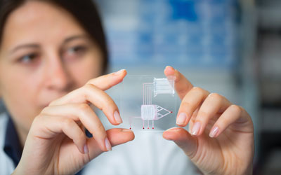
Lab-On-A-Chip and microfluidics
Lab-On-A-Chip technologies are based on an automated, micro- or nano-scale laboratory that enables sample preparation, fluid handling, and analysis and detection steps to be carried out within the confines of a single microchip. The technology is based on microfluidics, and the most familiar consumer application is ink-jet printing. Microfluidics allows for the manipulation of minute amounts of liquid in miniaturised systems that are composed of a network of channels and wells that are etched onto glass or polymer chips. Pressure or voltage gradients move pico or nanolitre volumes through the channels in a well-controlled manner that enables sample handling, mixing, dilution, electrophoresis and chromatographic separation, staining and detection. Currently available lab chips analyse protein, DNA, RNA and whole cells in fluid samples.
One example of this type of technology utilises a microfluidics chip to separate rep-PCR amplicons for the purpose of identifying microorganisms. The system targets short, repeating sequences of unknown function that occur randomly throughout the DNA of an organism. Primers bind to these sequences, resulting in multiple fragments (i.e., the amplicons) of various lengths, which are subsequently added to the chip and separated by size and charge. The amplicons then pass through a laser, causing fluorescence of an intercalating dye. A unique rep-PCR fingerprint profile is created containing multiple bands of varying sizes and intensities, which is then compared with an internal database of similar profiles. If a match is found, a microbial identification is provided.
Microscale impedance detection
Purdue University has recently developed an impedance detection system that is composed of fine channels and chambers that have been etched onto a crystalline silicon substrate. Platinum electrodes measure impedance changes of very small volumes of micro – biological medium when microorganisms are present (this is the same concept as the benchtop impedance system I discussed in the first article of the year). Actually, the incubation and measurement chamber volume is less than one nanolitre. Therefore, the smaller the volume that is analysed, the faster the systems can detect changes in electrical impedance, conductance and/or capacitance of the medium.
Microarrays
Microarrays (also known as biochips or DNA chips) are collections of miniaturised test sites, arranged on solid substrates that permit many tests to be performed at the same time. They are composed of an orderly arrangement of protein or thousands of DNA or RNA fragments on glass, silicon or nylon substrates. This technology evolved from Southern blot technology, in which fragmented DNA is attached to a substrate and then probed with a known gene or DNA fragment, using fluorescent tags to allow visual detection. Microarrays are usually fabricated using a variety of technologies, including printing with fine-pointed pins onto glass slides, photolithography, ink-jet printing or electrochemistry. Other methods may also be used, such as in situ synthesis, whereby the probes are synthesised directly on the chip instead of spotting them on the array.
Applications for microarrays include nucleic acid sequence identification and measuring expression levels of genes. For example, the entire human genome (50,000 known genes and gene variants) has now been placed onto a single chip, and other microarray systems contain probes that can rapidly detect influenza A and Avian H5N1 (Bird Flu) strains.
One currently available system is now being used to identify up to 40 species of Mycoplasma in a microarray format. DNA is first extracted from the Mycoplasma culture and PCR is performed using primers specific for conserved and species-specific regions of the 16S-23S rRNA intergenic transcribed spacer (ITS) of the Mycoplasma DNA. The fluorescently labelled fragments are then hybridised to the microarray chip. The chip contains probes for both species-specific targets and a universal probe for all Mycoplasmas, and if one of the targets is present, the system will provide a positive response.
Biosensors
Biosensors can detect an analyte that is comprised of a biological component combined with a physicochemical detector component. Nineteenth-century coal miners used a canary in a cage as a primitive biosensor to detect lethal concentrations of gas. Today, the most widespread example of a commercially available biosensor is the blood glucose monitor, but future uses will include remote sensing of airborne bacteria and detection of pathogens. The biological component of a biosensor may contain tissue, microorganisms, organelles, cell receptors, enzymes, antibodies, nucleic acids or whole cells. The detector element works in a physicochemical manner and may include optical, electrochemical, thermometric, piezoelectric or magnetic technologies. A transducer associates both the biological and the detector components. One example of a currently available biosensor performs immunological assays, where the components are spotted in one-micron tall wells using less than one nanolitre of material. In this example, biosensors have been used to rapidly detect the presence of influenza. It is probable that we will see future designs for the detection of other microorganisms, including those associated with pharmaceutical product and environmental contamination.
Combination systems
Combining multiple MEMS platforms results in a hybrid system that is superior to the individual platforms themselves. An example is when we combine microfluidics and microarrays. One technology that is currently used in the diagnostics field provides a fully integrated PCR reactor and a microarray used to hybridise and detect PCR amplicons. The technology’s chip is mounted on a 1×3 inch plastic slide that provides the necessary mechanical, thermal, electrical and fluidic connections. When a test sample size of 2-8 μl is used, the PCR reactions are three times faster than conventional thermocyclers, and a portable, customised fluorescent-based optical reader analyses the microarray in only a few seconds. The system hosts a pathogen panel to identify 10 sepsiscausing bacterial species as well as methicillinresistant strains of Staphylococcus aureus from contaminated blood culture samples.
Nanotechnology
Nanotechnology operates at the atomic, molecular, or macromolecular range of approximately 1-100 nanometres to create and use structures, devices, and systems that have novel properties. The key to this technology is high-voltage electron-beam (or E-beam) lithography, in which a beam of electrons is scanned across a surface covered with a thin film, called a resist. The electrons produce a chemical change in the resist, which allows the surface to be patterned.
Nanotechnology has allowed us to fabricate nanoarrays, with molecules placed at defined locations on a surface with nanometre spatial resolution. Nanoarray spots can include biological samples such as proteins, DNA, RNA and whole viruses, as well as non-biological samples such as chemical solutions, colloids and particle suspensions, and are the next evolutionary technological step in the miniaturisation of bioaffinity tests for proteins, nucleic acids and receptor-ligand pairs. These arrays utilise approximately 1/10,000th of the surface area occupied by a conventional microarray, and over 1,500 nanoarray spots can occupy the area required for a single microarray spot.
One nanoarray system prints biological and non-biological materials onto silicon chips and other surfaces with ultra-micro spot sizes ranging from 1 – 20 μm, and in the nanometre range to 250 nanometres. The technology utilises surface patterning tools, or SPTs, which are microcantilever-based microfluidic handling devices. The droplet volumes these microcantilevers deliver to the nanoarray are in the femtolitre and attolitre range (10-12 millilitres and 10-15 millilitres, respectively). Nanoarrays offer a number of advantages, including labelfree detection (via atomic force microscopy). They work in both solutions and biological liquids, retaining biological activity for subsequent analyses.
Microcantilevers and nanocantilevers
In the previous section, I mention the use of microcantilevers to deliver very small droplet volumes to the surface of a nanoarray. A microcantilever is a device that can also act as a physical, chemical or biological sensor by detecting changes in cantilever bending or vibrational frequency. It can be considered as the miniaturised counterpart of a diving board that moves up and down at a regular interval based on mass. The mass changes when contaminants land on the devices, causing them to vibrate at a different ‘resonant frequency’, which can be quickly detected. The sizes of micro – cantilevers are in the micron range and can be fabricated in different shapes. Scientists at the University of California at Berkeley have recently developed a chip-level microcantilever array that is expected to provide a quantitative, label-free and low-cost platform for the detection of various biomolecules, such as DNA and proteins. But we can go even smaller than that. Scientists at Purdue University have developed advanced sensors capable of detecting minute quantities of viruses, bacteria and other contaminants in air and fluids by coating nanocantilevers with proteins and antibodies that attract the contaminants. The applications for such devices include use in hospital environmental monitoring and homeland security (e.g., the detection of weaponised biological particles). Thousands of these nanocantilevers can be fabricated on a 1 cm2 chip, and each nanocantilever is only a few microns in length and 20 nanometres in thickness.
Summary
MEMS represent the next generation in microorganism detection platforms, reducing our instrumentation footprint to the micro and even the nanotechnology level. We are already using these technologies to detect certain types of contaminants, such as Mycoplasma, but I envision their future use in manufacturing as Process Analytical Technology (PAT) platforms, where chips may be used in future hand-held devices, or embedded within product and process streams for the continuous, real-time monitoring and detection of microorganisms, thereby completely eliminating the need for offline assays. These are truly exciting times for rapid methods, and they will only get better!
For additional references on these and other types of RMMs, please visit the Technologies and Reference pages on my educational rapid methods website, http://rapidmicromethods.com. My blog has also discussed recent advances in MEMS technologies, and you can follow the new technology discussions at http://blog.rapidmicromethods.com/.
About the Author
Dr. Michael J. Miller is an internationally recognised microbiologist and subject matter expert in pharmaceutical microbiology and the design, validation and implementation of rapid microbiological methods. He is currently the President of Microbiology Consultants, LLC (http://microbiologyconsultants.com). For more than 23 years, he has held numerous R&D, manufacturing, quality, and consulting and business development leadership roles at Johnson & Johnson, Eli Lilly and Company, Bausch & Lomb, and Pharmaceutical Systems, Inc. In his current role, Dr. Miller consults with multinational companies in providing technical, quality and regulatory solutions in support of RMMs, sterile and non-sterile pharmaceutical manufacturing, contamination control, isolator technology, validation and microbiological PAT. He also provides comprehensive training for his clients in the areas of rapid method validation and implementation.
Dr. Miller has authored over 100 technical publications and presentations in the areas of rapid microbiological methods, PAT, ophthalmics, disinfection and sterilisation, is the editor of PDA’s Encyclopedia of Rapid Microbiological Methods, and is the owner of http://rapidmicromethods.com, a website dedicated to the advancement of rapid methods. He currently serves on a number of PDA’s program and publication committees and advisory boards, is co-chairing the revision of PDA Technical Report #33: Evaluation, Validation and Implementation of New Microbiological Testing Methods, and routinely provides RMM training programs for the industry and professional organisations worldwide.
Dr. Miller holds a Ph.D. in Microbiology and Biochemistry from Georgia State University (GSU), a B.A. in Anthropology and Sociology from Hobart College, and is currently an adjunct professor at GSU. He was appointed the John Henry Hobart Fellow in Residence for Ethics and Social Justice, awarded PDA’s Distinguished Service Award and was named Microbiologist of the Year by the Institute of Validation Technology (IVT).