MitoCheck: checking mitosis
Posted: 20 July 2006 | | No comments yet
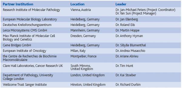
MitoCheck is a multi-national, multi-disciplinary research project on cell cycle control. It is funded by the European Union within its 6th framework program (FP6). Leading scientists from 11 research institutes, universities and industry in Austria, Germany, UK, Italy and France with a wide range of expertise in molecular and cell biology, biochemistry, modern microscopy techniques, proteomics, bio-informatics and clinical pathology have joined forces to take on the challenge of unraveling the mystery of cell division using RNAi.
MitoCheck is a multi-national, multi-disciplinary research project on cell cycle control. It is funded by the European Union within its 6th framework program (FP6). Leading scientists from 11 research institutes, universities and industry in Austria, Germany, UK, Italy and France with a wide range of expertise in molecular and cell biology, biochemistry, modern microscopy techniques, proteomics, bio-informatics and clinical pathology have joined forces to take on the challenge of unraveling the mystery of cell division using RNAi.
MitoCheck is a multi-national, multi-disciplinary research project on cell cycle control. It is funded by the European Union within its 6th framework program (FP6). Leading scientists from 11 research institutes, universities and industry in Austria, Germany, UK, Italy and France with a wide range of expertise in molecular and cell biology, biochemistry, modern microscopy techniques, proteomics, bio-informatics and clinical pathology have joined forces to take on the challenge of unraveling the mystery of cell division using RNAi.
The proliferation of cells depends on the replication of their genomes in S-phase of the cell cycle and on segregation of the replicated genomes during mitosis. The latter is an immensely complex process that involves events such as dissolution of the nuclear membrane, changes in chromosome organisation and re-organisation of the spindle apparatus. Mistakes during mitosis contribute to cancer whereas mistakes during meiosis are the leading cause of infertility and mental retardation.
During past decades, a great deal has been learnt about how individual genes function in certain aspects of mitosis. How cells coordinate many disparate but inter-locking processes during mitosis is, however, poorly understood. One of the well-established facts is that protein kinases such as cyclin-dependent kinase 1 (Cdk1), Polo-like kinase 1 (Plk1) and Aurora kinases play fundamental roles in mitosis (Nasmyth, 2001). Nevertheless, the actual molecular functions of these enzymes remain largely mysterious. The main objective of the Integrated Project MitoCheck is to understand how mitotic kinases orchestrate the many events of mitosis. In particular, MitoCheck wants to identify all the proteins that are required for mitosis, analyse which of these are phosphorylated by mitotic kinases and begin to study how phosphorylation changes the activity of these mitotic proteins.
In the past, identification of kinase substrates has been hampered by difficulties in mapping phosphorylation sites, in experimentally controlling protein kinase activity, and in evaluating the physiological consequences of defined phosphorylation sites. MitoCheck has been founded because all these hurdles can now principally be overcome by new technologies, namely the use of RNA interference (RNAi) to identify in a systematic, genome-wide manner potential substrates, affinity tagging to purify protein complexes, the use of small molecule chemicals to inhibit specific kinases in a controlled fashion and mass spectrometry to identify phosphorylation sites on complex subunits.
As the first step to systematically search for substrates of mitotic kinases, MitoCheck is carrying out genome-wide RNAi screens in human cells to identify human genes required for mitosis (Neumann et al., 2006). A genome-wide collection of synthetic small interfering RNAs (siRNAs) is introduced into human cells via solid-phase transfection to deplete all 22,000 human genes one by one. The behaviour of the cells after transfection is then recorded in real time by live cell video microscopy during a period of time that normally spans several cell cycles. All video microscopy movies generated in the screen will then be analysed with the help of the automated image analysis tools developed by MitoCheck partners. So far 177 000 movies have been recorded (corresponding to 42 terabytes of data), and once their analysis and annotation has been completed all of these will in the future be displayed on the MitoCheck Web site (www.mitocheck.org). This dataset includes a wide range of cellular phenotypes, mitotic and non-mitotic. The MitoCheck database will therefore be a unique and highly valuable source of information, not only for the cell cycle field but also for many other areas in the life sciences, ranging from cell biology to genomics/proteomics and basic medical research areas.
A subset of the proteins that are crucial for mitosis is currently being tagged with green fluorescent protein (GFP) and with an affinity purification tag. Genes encoding the tagged proteins will then be stably expressed from their own regulatory elements by using bacterial artificial chromosomes (BACs) in human cell lines. The GFP tag will allow visualisation of the subcellular localisation of these proteins during cell cycle, whereas the affinity tag will enable the identification of binding partners of these proteins via affinity purification followed by mass spectrometry. Since the phosphorylation state of these proteins and their binding partners are thought to be crucial for their function during mitosis, much of MitoCheck’s efforts will be devoted to identifying the mitosis-specific phosphorylation sites on them. For this purpose, MitoCheck has established mass spectrometry methods for the rapid identification of phosphorylation sites on the subunits of mitotic protein complexes (Herzog et al., 2005). Identifying the kinase(s) that are responsible for the generation of certain phosphorylation sites in vivo is a big challenge. MitoCheck is taking two approaches to tackle this difficult problem. First, cells are pretreated with specific chemical inhibitors of mitotic kinases (Hauf et al., 2003; Strebhardt and Ullrich, 2006) before the proteins are purified and analysed by mass spectrometry to explore whether a given phosphorylation site depends on the activity of a particular kinase. In parallel, MitoCheck is creating alleles of mitotic kinases whose activities can be controlled by synthetic analogs of ATP (named after their inventor ‘Shokat analogs’). These alleles will help to identify the potential substrates of mitotic kinases. In addition, MitoCheck is using protein crystallography techniques to understand how existing kinase activators or inhibitors interact with their target kinases. MitoCheck has already succeeded in co-crystallising the mitotic kinase Aurora B with a fragment of its activator INCENP and its small molecule inhibitor Hesperadin (Sessa et al., 2005).
Some mitotic kinases are over-expressed in human tumors. One important objective of MitoCheck is to develop assays to systematically evaluate the clinical utility of mitotic kinases as diagnostic or prognostic markers, and to identify tumor types as potential targets for small molecule inhibitors of the respective kinase. MitoCheck has already developed clinical assays for expression levels of mitotic kinases in human tumors and has initiated first clinical trials on cancer patients with these new assays.
Understanding a process as complex as mitosis is not an easy task. Project coordinator Jan-Michael Peters at the Research Institute of Molecular Pathology in Vienna is nevertheless optimistic: “We have set very ambitious goals, which no single research partner could have tackled alone. By bringing together a group of excellent European scientists who contribute expertise in rather diverse areas, we can hope to solve a complex biological puzzle.”


References
Nasmyth K. 2001. A prize for proliferation. Cell. 107(6), 689-701.
Neumann B, Held M, Liebel U, Erfle H, Rogers P, Pepperkok R, Ellenberg J. 2006. High-throughput RNAi screening by time-lapse imaging of live human cells. Nat Methods. 3(5), 385-90.
Herzog F, Mechtler K, Peters JM. 2005. Identification of cell cycle-dependent phosphorylation sites on the anaphase-promoting complex/cyclosome by mass spectrometry. Methods Enzymol. 398, 231-45.
Hauf S, Cole RW, LaTerra S, Zimmer C, Schnapp G, Walter R, Heckel A, van Meel J, Rieder CL, Peters JM. 2003. The small molecule Hesperadin reveals a role for Aurora B in correcting kinetochore-microtubule attachment and in maintaining the spindle assembly checkpoint. J Cell Biol. 161(2), 281-94.
Strebhardt K, Ullrich A. 2006. Targeting polo-like kinase 1 for cancer therapy. Nat Rev Cancer. 6(4),321-30.
Sessa F, Mapelli M, Ciferri C, Tarricone C, Areces LB, Schneider TR, Stukenberg PT, Musacchio A. 2005 Mechanism of Aurora B activation by INCENP and inhibition by hesperadin. Mol Cell. 18(3), 379-91


