PEG-IONCs show potential as next-generation MRI contrast agents
Posted: 31 July 2017 | Dr Zara Kassam (European Pharmaceutical Review) | No comments yet
A research team formed have tested a new non-toxic contrast agent for magnetic resonance imaging (MRI) and magnetic resonance angiography (MRA) that could be superior to the current dye, gadolinium.
Previously, the Institute for Basic Science (IBS) team designed ultra-small T1 iron oxide nanoparticles (PEG-IONCs), proving the possibility to synthesise them in large quantities. Their research has now leaped forward: “Research on mice cannot directly translate to humans, so we wanted to test if these nanoparticles work on large animals, like dogs, rabbits and monkeys. Eventually, our goal is to be able to understand if they can become a new diagnostic tool for humans,” comments Hyeon Taeghwan, director of the Center for Nanoparticle Research.
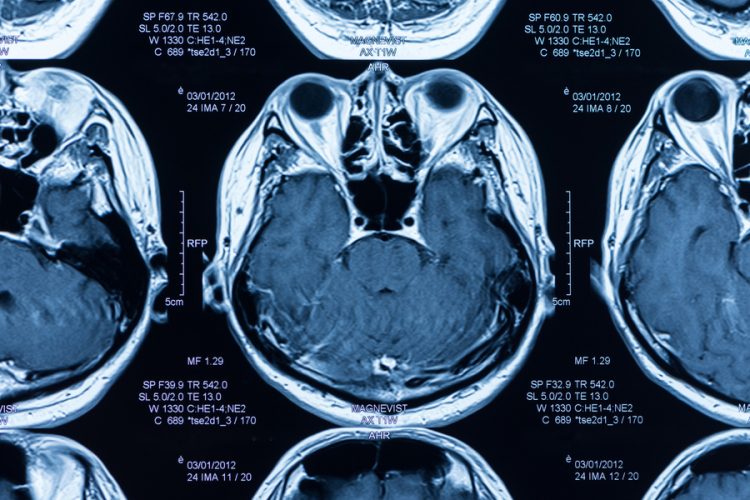
Mr Taeghwan explains: “Let’s take the example of a MRI analysis of a brain with Alzheimer’s: iron oxide in the blood vessels would appear as black and the amyloid plaques as gray. It is very hard to recognise the plaques from the background. For it is reason, the current iron oxide nanoparticles are not used anymore and we started to look for other options.” This occurs because gadolinium is a so-called T1-type contrast agent, while the current iron oxide is classified as T2.
With a small diameter and an non-harmful coating, the PEG-IONCs boast several desirable features. PEG-IONCs’ hydrodynamic diameter of about 12 nanometers is much smaller and more uniform than commercially available iron oxide nanoparticles. Moreover, in preparing IONCs, IBS scientists used safe components, such as oleic acid, oleyl alcohol, and polyethylene glycol (PEG), that are commonly employed in pharmaceutical formulations.
Hematological and tissue compatibility studies revealed that PEG-IONCs are highly biocompatible. To give an example, most of the clinically available iron oxide-based MRI contrast agents, such as ferumoxide and ferumoxtran, have to be slowly infused to minimise the incidence of hypotension and other severe side effects. On the other hand, PEG-IONCs were administered trouble-free, like gadolinium.
Finally, after the successful use of PEG-IONCs in static MRA, the scientists conducted a more challenging dynamic imaging to see vascular flow patterns of cerebral ischemia in dogs and monkeys. In the experiment, images were taken at several time points, every 1.5 seconds after injection of the contrast agent to see the blood perfusion patterns in the brain. An occlusion in the middle cerebral artery was detected.
While further rigorous preclinical investigations are required, the current pilot studies on nonhuman primates clearly demonstrate a great potential of PEG-IONCs for next-generation T1 MRI contrast agent.
Related topics
Related organisations
Center for Nanoparticle Research, Institute for Basic Science