

Non-invasive technique for probing cells may reveal disease
Posted: 3 August 2017 | Dr Zara Kassam (European Pharmaceutical Review) | No comments yet
Engineers have devised a non-invasive way to assess a cell’s mechanical properties simply by observation…
Engineers have devised a non-invasive way to assess a cell’s mechanical properties simply by observation, the researchers use standard confocal microscopy to zero in on the constant, jiggling motions of a cell’s particles — telltale movements that can be used to decipher a cell’s stiffness. Unlike optical tweezers, the team’s technique is non-invasive, running little risk of altering or damaging a cell while probing its contents.
Determining the mechanical properties of cells may thus help doctors diagnose and track the progression of certain diseases. Current methods for doing this involve directly probing cells with expensive instruments, such as atomic force microscopes and optical tweezers, which make direct, invasive contact with the cells.
“There are several diseases, like certain types of cancer and asthma, where stiffness of the cell is known to be linked to the phenotype of the disease,” says Prof Ming Guo, the Brit and Alex d’Arbeloff Career Development Assistant Professor in MIT’s Department of Mechanical Engineering. “This technique really opens a door so that a medical doctor or biologist, if they would like to know the material property of cell in a very quick, noninvasive way, can now do it.”
Prof Guo surmised that there might be a way to tease out temperature-driven motions in a cell by looking at the cell within a very narrow time-frame. They realised that particles energised solely by temperature exhibit a constant jiggling motion. No matter when you look at a temperature-driven particle, it’s bound to be moving.
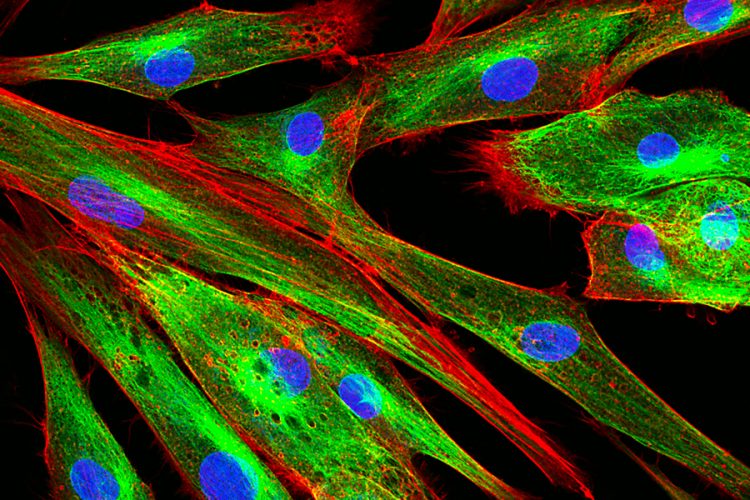
In contrast, active processes that can knock a particle around a cell’s cytoplasm do so only occasionally. Seeing such active movements, they hypothesised, would require looking at a cell over a longer time-frame. To test their hypothesis, the researchers carried out experiments on human melanoma cells. They injected small polymer particles into each cell, then tracked their motions under a standard confocal fluorescent microscope. They also varied the cells’ stiffness by introducing salt into the cell solution — a process that draws water out of cells, making them more compressed and stiff.
The researchers recorded videos of the cells at different frame rates and observed how the particles’ motions changed with cell stiffness. When they watched the cells at frequencies higher than 10 frames per second, they mostly observed particles jiggling in place; these vibrations appeared to be caused by temperature alone. Only at slower frame rates did they spot more active, random movements, with particles shooting across wider distances within the cytoplasm.
Within a cell, organelles such as mitochondria and lysosomes are constantly jiggling in response to the cell’s temperature. However, Prof Guo said, there are also “many minispoons” stirring up the surrounding cytoplasm, in the form of proteins and molecules that, every so often, actively push vibrating organelles around like billiard balls.
The constant blur of activity in a cell has made it difficult for scientists to discern, simply by looking, which motions are due to temperature and which are due to more active, “spoon-like” processes. This limitation, Prof Guo said, has “basically shut the door on using Einstein’s equation, that makes it possible to calculate a material’s mechanical properties by observing and measuring the movement of particles in that material, and pure observation to measure a cell’s mechanical properties
The team’s results suggest that if researchers observe cells at fast enough frame rates, they can isolate particle motions that are purely driven by temperature, and determine their average displacement — a value that can be directly plugged into Einstein’s equation to calculate a cell’s stiffness.
“Now if people want to measure the mechanical properties of cells, they can just watch them,” Prof Guo says.
The team is working with doctors at Massachusetts General Hospital, who hope to use the new, noninvasive technique to study cells involved in cancer, asthma, and other conditions in which cell properties change as a disease progresses.
“People have an idea that structure changes, but doctors want to use this method to demonstrate whether there is a change, and whether we can use this to diagnose these conditions,” Prof Guo said.
Related topics
Related organisations
Massachusetts General Hospital, MIT's Department of Mechanical Engineering
