3d simulation gives authentic representation of left-hand heart mechanisms
Posted: 2 March 2018 | European Pharmaceutical Review | No comments yet
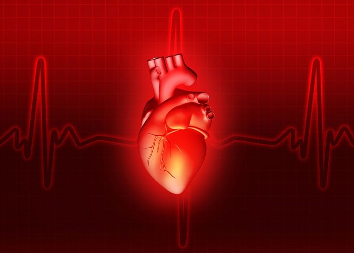
A team of scientists in Italy has developed a 3d model that uniquely presents an holistic demonstration of how the left-hand chambers of the heart interact with blood flow and its component parts. The ultimate aim is better prevention and treatment of cardiac conditions.


Understanding how blood flow in the left-hand side of the heart affects the way it works could be the key to preventing cardiac problems and this is the raison d’être of the 3d model.
In a new study, Valentina Meschini from the Gran Sasso Science Institute, L’Aquila, Italy and colleagues introduce a model that examines the mutual interaction of the blood flow with the individual components of the heart. Their work uniquely offers a more holistic and accurate picture of the dynamics of blood flow in the left ventricle. The authors also perform some experimental validations of their model.
3d awareness of heart pressures
The left side of the heart is the part that is most vulnerable to cardiac problems – particularly the left ventricle, which has to withstand intense pressure differences and is therefore under the greatest strain. As a result, people often suffer from valve failure or impairment of the muscle tissue, known as myocardium.
In their 3d numerical simulation study, the authors develop a mathematical model that accounts for the fact that parts of the left side of the heart – including the left ventricle, the mitral valve and the chordae tendineae (tendinous chords) – are coupled in a twofold manner with the blood flowing through the heart. The mitral valve has two flaps and lies between the left atrium and the left ventricle, while the tendinous chords connect the heart muscles to the heart valves.
Until now, most cardiac models have considered separate components of the heart – either the ventricle or the mitral valve – but they have never approached the whole combination as a synergistic system. Another key shortcoming of previous models was their failure to take account of either the interaction between the blood and the heart structure, which can lead to deformation of the heart, or the structure of the heart chambers under the load of the passing blood flow.
Commenting on the unique development of their 3d model, Meschini said: “The inclusion of a chorded mitral valve in an already complex system like the left ventricle is a challenging step forward to an uncompromised computational model of the heart,”
The authors conclude that the effects of the tendinous chords on the mitral valve are more complex than initially assumed. They also reveal the importance of the effects of blood dynamics and a different type of ventricle deformation caused by the pulling action of said chords on the myocardium.
“The next step in this work would be to replace the imposed flow-rate with an active contraction/relaxation of the ventricle. This could be achieved by coupling the fluid-structure interaction model with an electrophysiology model able to provide the propagation of the electrical stimulus through the whole ventricle. This additional feature will make the computational model much more realistic and reliable,” explains Meschini
Meschini adds that the final goal is to simulate the whole heart so that the right ventricle and the two atriums in the system work synergistically.
“We believe that coupling this with the electrophysiology model would be key, and would give us a reliable tool which can be used for virtual checks, to test new medicine or different intervention measures and avoid in vivo experiments with animals or real patients.”
This research was published in the European Physical Journal E.


