Nano-optic endoscope penetrates deep tissue at high resolution
Posted: 7 August 2018 | European Pharmaceutical Review | No comments yet
The images captured by the nano-optic endoscope shows cellular structures in fruit flesh clearly, and tissue layers and fine glands in sheep…
Diagnosing many diseases relies on biopsies of internal tissue collected from the affected region. In current practises, the use of endoscopic imaging techniques and conventional optical elements to access hard-to-reach areas have a high error rate because of the inability of current technology to visualise disease sites accurately.
Researchers at Massachusetts General Hospital (MGH) and pioneers of flat metalens technology at the Harvard John A. Paulson School of Engineering and Applied Sciences (SEAS) have collaborated to develop a new type of endoscopic imaging catheters. They have been given the term ‘nano-optic endoscopes’, and the researchers aim to overcome the limitation of current systems.
“Clinical adoption of many cutting-edge endoscopic microscopy modalities has been hampered due to the difficulty of designing miniature catheters that achieve the same image quality as bulky desktop microscopes,” said Assistant Professor Melissa Suter at MGH and Harvard Medical School (HMS).
“The use of nano-optic catheters that incorporate metalenses into their design will likely change the landscape of optical catheter design, resulting in a dramatic increase in the quality, resolution, and functionality of endoscopic microscopy. This will ultimately increase clinical utility by enabling more sophisticated assessment of cell and tissue microstructure in living patients.”
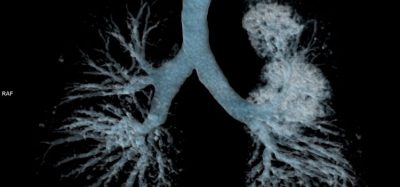
As a trial run, the team used the nano-optic endoscope to image the flesh of fruit, swine and sheep airways and human lung tissue. The researchers showed that the nano-optic endoscope can image deep into tissue at significantly higher resolutions than current catheter imaging options.
“Metalenses based on flat optics are a game changing new technology because the control of image distortions necessary for high resolution imaging is straightforward compared to conventional optics, which requires multiple complex shaped lenses,” said Professor of Applied Physics and Vinton Hayes Senior Research Fellow in Electrical Engineering at SEAS, Dr Federico Capasso.
The images captured by the nano-optic endoscope shows cellular structures in fruit flesh clearly, along with tissue layers and fine glands in sheep and swine. In human lung tissue, the researchers were able to clearly identify structures that corresponded to irregular glands, indicating the presence of adenocarcinoma.
“The versatility and design flexibility of the nano-optic endoscope significantly elevates endoscopic imaging capabilities and will likely impact diagnostic imaging of internal organs,” said Dr Hamid Pahlevaninezhad, Instructor in Medicine at MGH and HMS.
“We demonstrated an example of such capabilities to achieve high-resolution imaging at greatly extended depth of focus.”
The team is confident that their approach will lead to a ‘class of optical systems’ and will have a broad range of applications in the vast area of science and technology. The researchers aim to explore other uses for the nano-optic endoscope and look to develop a polarisation-sensitive nano-optic endoscope, which could contrast between smooth muscle, blood vessels and collagen.
The research has been published in Nature Photonics.