Combined MRI and SPECT imaging could guide ventricular tachycardia
Posted: 31 January 2019 | Iqra Farooq (European Pharmaceutical Review) | No comments yet
A study has found that a combination of imaging techniques could guide and improve current ablation strategies used to treat patients…
Research has shown that adding functional imaging to structural imaging of patients with ventricular tachycardia (VT) could improve current ablation strategies.
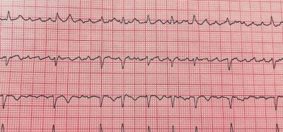
When a team of researchers combined cardiac magnetic resonance imaging with iodine-123 metaiodobenzylguanidine (123I-MIBG) SPECT imaging, it helped to identify specific subsets of heart tissue that is more prone to arrhythmia. Ventricular arrhythmias, also known as abnormal heartbeats, originate from the bottom chambers of the heart, and are one of the main causes of sudden cardiac death.
In the US, ventricular arrhythmias is responsible for around 300,000 deaths each year. Ablation of VT is a known proven treatment for individuals who have a history of heart attacks. Ablation is a treatment that looks to control or correct certain abnormal heart rhythms by breaking the abnormal circuits in the heart, and destroying the areas of the heart muscle that trigger arrhythmias.
Currently, the initial challenge has been the identification of the area responsible of arrhythmia.
“The amount of scar tissue can often account for more than half of the left ventricle myocardium,” noted Dr Timm Dickfeld, Director of electrophysiology research at the University of Maryland School of Medicine. “Ablating such a large amount of the myocardium is often not desirable and very time-intensive.”
During the study, 15 patients with ischemic cardiomyopathy who were scheduled for radiofrequency ablation for drug-refractory VT were followed. Cardiac MRI and 123I-MIBG SPECT was carried out on each patient, along with high resolution bipolar voltage mapping.
These techniques assessed various adaptations found in VT: abnormal innervation, tissue scarring and low-voltage area. Results were then compared with each other to determine which of them were present in the affected area of the heart tissue.
“Results from this study show that nuclear medicine can be used to develop novel, cutting-edge strategies for risk stratification and arrhythmia treatment,” said Dr Dickfeld. “The study highlights the importance of close collaboration among the nuclear medicine, radiology and electrophysiology departments, which we are lucky to have at the University of Maryland, to move the field forward.
“We hope that we can build on the insights provided by this research to develop new treatment algorithms that will result in potentially shorter and more efficient treatment of patients with ventricular arrhythmias.”
The study was published in The Journal of Nuclear Medicine.
Related topics
Analytical techniques, Clinical Development, Clinical Trials, Drug Development, Research & Development (R&D)