Biochemical fingerprints reveal diabetes progression
Posted: 22 August 2017 | Dr Zara Kassam (European Pharmaceutical Review) | No comments yet
Researchers have described a new method to study biochemical changes that occur in the pancreas during the development of diabetes…
Researchers have described a new method to study biochemical changes that occur in the pancreas during the development of diabetes.
The method is based on molecular spectroscopy and can be used to extract biochemical profiles containing information about disease progression. The method could facilitate improved understanding of the mechanistic processes on molecular and cellular levels that are key to the development of diabetes.
The method uses vibrational micro-spectroscopic technology, including Fourier Transform Infrared (FT-IR) and Raman micro-spectroscopy. By using advanced statistical methods, researchers can filter out noise. This results in a better overview and allows researchers to focus on the important factors.
“This method is well-suited for studying biological samples, since it does not damage the sample, does not require external markers such as antibody labels, and can be used in microscopy settings. The method can for example be used to determine which cell types are affected in a certain tissue, where and how,” said András Gorzsás, researcher at the Department of Chemistry and co-author of the study.
The researchers describe how a method for multivariate statistical analysis enables them to handle multiple variables simultaneously and thus analyse complex data from vibrational microspectroscopy of the pancreas.
Using this method, which until now has been used primarily to study plant tissues, the researchers show that it is possible to discover previously unknown biochemical changes in the pancreas during disease development. In addition, previously known changes in the tissue may also be detected, but at even earlier stages of disease progression compared to what has been described by other techniques.
“By using this method we can create biochemical fingerprints of all changes occurring in the pancreas. The fingerprints inform us of what cell type we are looking at, which animal model it comes from and how far the disease has progressed. These fingerprints are so precise that even unknown samples can be classified if there is available reference material,” said Ulf Ahlgren, Professor of Molecular Medicine and co-author of the article.
The method can be used to analyse both mice and human pancreas from the outside of the organ, i.e. without the need to obtain tissue samples. Moreover, the researchers demonstrate in a transplantation experiment that pancreatic tissue may be studied in. In addition to studying mechanistic aspects of diabetes development and manifestation, the researchers hope that the method can be used to develop better prognostic and diagnostic tools for diabetes.
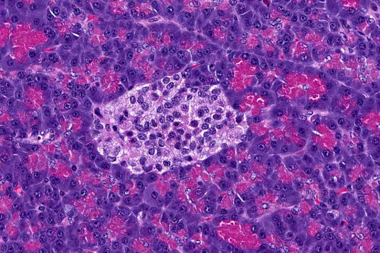
“I believe this possibility to study pancreatic tissue and especially the biochemistry of the insulin-producing Islets of Langerhans in the living organism is a very interesting opportunity for diabetes research. The method could prove useful for example to study the direct effects of anti-diabetic therapies on the biochemical composition and function of insulin-producing cells,” said Prof Ahlgren.
The researchers from the Umeå University in Sweden are also hopeful that their findings can lay the foundations for developing better tools for identifying cancer tissue to be surgically removed as part of pancreatic cancer treatment. The study was a collaborative effort with researchers at NTNU in Trondheim, Norway, and Karolinska institutet. The research was made possible by funding from the Swedish Research Council and the Kempe Foundations.