Accelerate food, pharmaceutical and particle analysis with Raman imaging
ABOUT THIS WEBINAR
This webinar describes how Raman microscopy can help solve some of the most vital and challenging questions that researchers face in 3D imaging and microparticle analyses of food, pharmaceuticals and microplastics, along with providing measurement examples from each field.
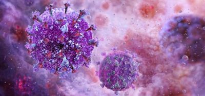
The session begins with a brief introduction to the principles of Raman microscopy and its associated hardware and software. Raman microscopy, which is based on the scattering of light by molecules or crystal lattices, can detect photons whose energies are shifted slightly from the excitation wavelength to serve as a unique “fingerprint” for each chemical component. The method is nondestructive and requires no dyeing or other specialised sample preparation. The ability to quickly and easily identify materials from small sample volumes or low concentrations makes it ideally suited to investigating microparticles.
Raman database management software will also be covered, as it can expedite the identification of investigated substances and allows newly acquired spectra to be saved for future reference.
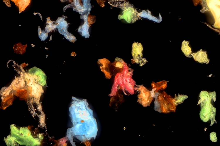
The use of Raman microscopy for automated particle analyses will then be described, with a focus on the finding, classifying and identifying of particle components. Investigations of this sort are especially important in establishing the purity of raw materials, such as water, used in the production of foods, drinks and pharmaceuticals. The varieties of packaging in which those products are distributed are also of interest as they influence the shelf life of their contents and are a potential source of microplastics in the environment. Employing the advantages of Raman microscopy to investigate particulate samples holds great promise in accelerating the workflow, refining the precision and amplifying the analytical power of such measurements.
Relevant application examples of the technique from food science, pharmaceutical research and microplastics research are presented, showing its versatility and speed.
After covering experimental procedures and demonstrating the capability, utility and accessibility of confocal Raman microscopy and automated particle analysis, we host a question and answer session to further explore the technique interactively.
Key learning points:
- Learn what Raman microscopy is, before being introduced to its operational principles and hardware considerations
- Discover how the technique can be useful for analyses in food and pharmaceutical research
- See how large numbers of microparticles and microplastics in a sample can be quickly found, classified and identified using automated routines
- Determine how to analyse drug delivery systems and food products.
KEYNOTE SPEAKERS
David Steinmetz, Director of Applications & Support, WITec GmbH

Dr Steinmetz obtained a Physics PhD from the Quantum Optics group of Professor Dr Christoph Becher at the University of Saarland in Saarbruecken, Germany. His work there was on single-photon sources based on single defect centres in solids. He joined WITec in 2011 and has been Director of Applications & Support since 2017.
Andrea Richter, Senior Applications Scientist, WITec GmbH

Andrea Richter holds a diploma degree in biology with a background in food technology and ecology. She has been a senior Applications Scientist at WITec since 2001.
Related topics
Analytical techniques, Environmental Monitoring, Microbiology, Near Infrared Spectroscopy (NIR), Personalised medicine, QA/QC, Raman Spectroscopy, Research & Development (R&D)