LC-TEM provides a window into the nanoscale
Posted: 26 April 2018 | European Pharmaceutical Review | No comments yet
Researchers create a device that allows more electron beam to pass through liquid samples…
A team of scientists and engineers have investigated the mechanisms that degrade sample quality in liquid cell transmission electron microscopy (LC-TEM). They developed an LC-TEM device that uses multiple windows and patterned features to explore the impacts of high-energy electron bombardment on nanoparticles and sensitive biological samples.
The team explains that transmission electron microscopy (TEM) relies on a high-energy beam of electrons that passes through a sample. Whether the sample is from a battery electrode or bacterial cells, the passing electrons will scatter in a specific way reflecting the sample’s atomic structure. In LC-TEM, materials can be examined in a native state allowing dynamic observations, but the samples are liquid or suspended in liquid and have to be tightly sealed to withstand the space-like vacuum of the instrument. There is a balance between ensuring the liquid doesn’t evaporate while providing enough viewing space for the electron beam to pass through.
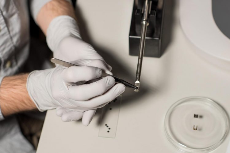
The research team create a tiny device that allows more of the microscope’s electron beam to pass through liquid samples. (Pacific Northwest National Laboratory).
“We have designed and fabricated new devices for holding liquid samples which give us more ‘window’ regions to collect images than were previously available,” says lead author, Trevor Moser. “Using these multiple windows, we were able to study how the history of electron irradiation influences the nucleation and growth of silver nanoparticles, the growth properties of which are sensitive to the radicals that are generated with the beam. We also used them to study how these radicals impact bacterial cells and demonstrate the extreme sensitivity of these biological samples to the electron beam.”
Irradiation from the high-energy beam used in LC-TEM can cause physical damage to samples. For example, the team found that when a cell was imaged–and was exposed to significant electron flux for the first time–observed nanoparticle movement relative to the cell membrane was a result of cell damage. That matters because the insight shows that the movement is an artefact of imaging the cell rather than watching cell dynamics happen in real-time.
“We were able to capture pristine images of cells using our multi-chamber device wherein the first image represented the cells first exposure to significant electron doses,” says James Evans, a senior scientist at PNNL.
“Since the native properties of the sample may be altered or changed by the effects of these electron beam-generated radicals,” says Tolou Shokuhfar, an Adjunct Professor of mechanical engineering at Michigan Tech “understanding the chemistry changes of a liquid sample as a result of electron irradiation is key to correct interpretation of data collected with this technique.”
As the differences of LC-TEM are gleaned, possible applications include gathering extremely high-resolution, detailed information on energy device and storage materials as well as disease detection, medical imaging and digging deep into the basics of cell activity. In terms of next steps, the team plans to focus on characterising more biological samples, which appear to be vulnerable to the effects of electron irradiation. The new LC-TEM device offers more windows into this complex atomic world, providing more chances for breakthroughs in energy and health.
The study has been published in Science Advances.
Related topics
Analytical techniques, Imaging, Material Identification, Microbial Detection, Microbiology, Microscopy