Application Note: Raman imaging in medical diagnosis and treatment
Posted: 14 December 2017 | WiTec | No comments yet
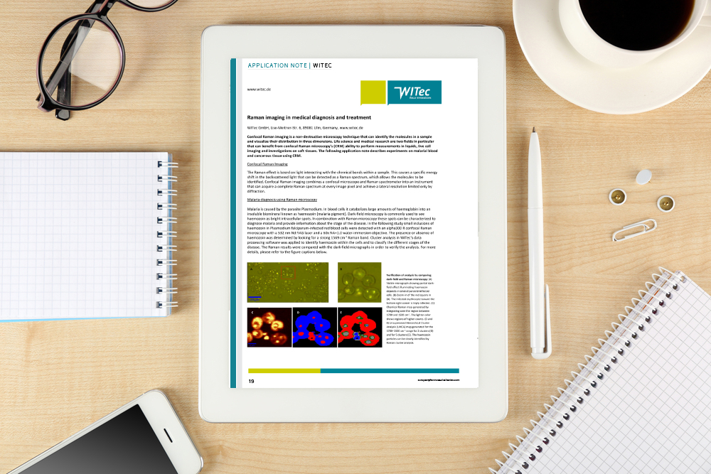
Confocal Raman imaging is a non-destructive microscopy technique that can identify the molecules in a sample and visualise their distribution in three dimensions…
Life science and medical research are two fields in particular that can benefit from confocal Raman microscopy’s (CRM) ability to perform measurements in liquids, live cell imaging and investigations on soft tissues.
The Raman effect is based on light interacting with the chemical bonds within a sample. This causes a specific energy shift in the backscattered light that can be detected as a Raman spectrum, which allows the molecules to be identified. Confocal Raman imaging combines a confocal microscope and Raman spectrometer into an instrument that can acquire a complete Raman spectrum at every image pixel and achieve a lateral resolution limited by diffraction.
The following application note describes experiments on malarial blood and cancerous tissue using CRM.
This whitepaper is restricted - login or subscribe free to access
 Thank you for visiting our website. To access this content in full you'll need to login. It's completely free to subscribe, and in less than a minute you can continue reading. If you've already subscribed, great - just login.
Why subscribe? Join our growing community of thousands of industry professionals and gain access to:
- bi-monthly issues in print and/or digital format
- case studies, whitepapers, webinars and industry-leading content
- breaking news and features
- our extensive online archive of thousands of articles and years of past issues
- ...And it's all free!
Click here to Subscribe today Login here
Related content from this organisation
- Application note: Analysing pharmaceutical tablets with confocal Raman imaging
- Raman spectroscopy: a useful tool in the fight against COVID-19
- Recent improvements in deep ultra-violet fluorescence devices for cleaning verification
- Application note: Combined Raman-SEM imaging for pharmaceutical studies
- QA/QC & Analytical Techniques In-Depth Focus 2020
Related topics
Cellular Imaging & Analysis (CIA), Imaging, Raman Spectroscopy, Research & Development (R&D), Spectroscopy